
A new imaging technique will improve cancer detection!
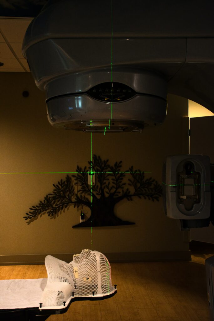
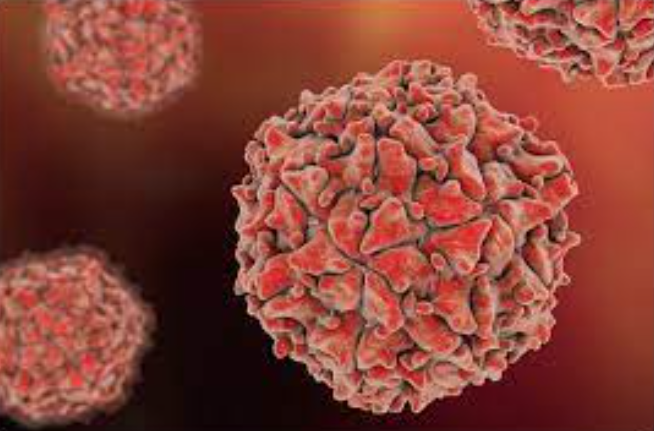
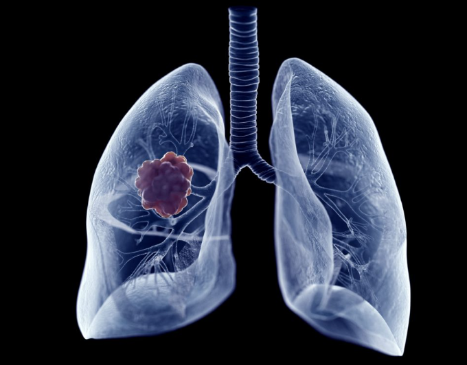
Currently the method used to detect cancer cells is positron emission tomography (particle that has the same electrical mass as the electron and the same electrical charge, but positive), these show the location of a tumor, generally based on the magnification of glucose intake, a hallmark of cancer cells.
Due to difficult economic access and regions that lack the necessary infrastructure to house and support the operation of this type of machines (PET), especially in developing countries and rural areas there has been an inevitable need for cheaper ways of imaging to improve the cancer care for a much larger population around the world.
Now, a team of researchers has begun testing a new technology that could provide a cheaper and faster imaging approach. This involves the use of specialized cameras to detect Cerenkov light, a faint glow naturally emitted by radioactive tracers. PET scans require these radioactive tracers to detect increased glucose metabolism in cancer cells. The new approach could make it possible to obtain critical information with most clinically used radioactive tracers, but without the PET machine, and for a fraction of the cost.
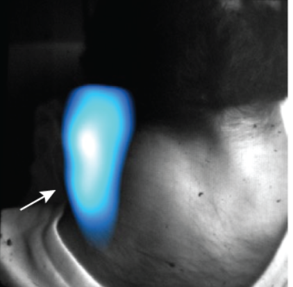
This method also provided information about whether their tumors were shrinking or otherwise responding to treatment. While it validates the technique’s potential as a cheaper and more portable imaging approach than PET, researchers are already looking for further improvements to make it more even more useful. This includes imaging of the whole body (rather than specific sites), imaging during an operation to help guide surgical decisions, and the use of a more sensitive camera to make image acquisition even faster and easier.
What do you think?


Responses