
Can you believe it? New three-dimensional map illuminates the “little brain” inside the heart

The heart has its own brain, now scientists have drawn a detailed map of this tiny brain, called the intracardiac nervous system, in rat hearts. The great boss of the heart is the brain, but the nerve cells of the heart also have something to say; These neurons are thought to play a crucial role in heart health, helping to adjust heart rhythms and perhaps protecting people against certain types of heart disease.
But until now, this local control system has not been mapped in great detail.To make their map, systems biologists imaged male and female rat hearts with a method called knife-edge scanning microscopy, creating detailed images of the heart’s anatomy. Those images could then be built into a three-dimensional model of the heart, the scientists also extracted individual neurons and measured the amount of gene activity within each cell.
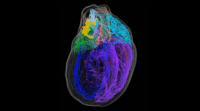
These measurements helped classify neurons in the heart into discrete groups; most of these groups of neurons are found at the top of the heart, where blood vessels enter and exit. Some of these clusters extended to the posterior part of the heart and were particularly abundant on the left side; With this new insight into individual groups, scientists can begin to study whether these groups have distinct jobs.
The tiny brain’s full three-dimensional map of the heart could ultimately lead to targeted therapies that could treat or prevent heart disease, scientists say.


Responses